As we spent time with family and friends over the holiday season, it became apparent that reading about Peanut’s condition and seeing it first-hand are two very different things. There were several good questions about some of the terms we’ve used in Peanut’s updates. It’s easy for us to explain what a broviac or a stoma is, but if you’ve never seen either, you likely wouldn’t be able to picture it in your mind. The whole month of August was like a refresher course of my high school anatomy class (which I passed with flying colors, but you wouldn’t have guessed it by some of the dumb questions I had for the doctors – I Googled more than I asked). 🙂
So we decided to take some pictures to better explain what’s going on with her. I have to admit, I think the reason we didn’t post any pictures earlier is because we were in a way still trying to get “comfortable” with seeing it all ourselves. Now it’s all second nature to us – it’s just all part of Peanut’s anatomy! We joke that it’s hard to remember what it’s like to have a baby without an ostomy bag. It just seems normal to us.
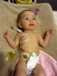
The thing we want our readers to keep in mind when you see these pictures, is that Peanut is not in any pain, nor does she have any discomfort – aside from being gassy! (no joke – it’s a common short bowel side affect) She is one happy little girl. 🙂
The Broviac:
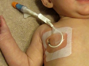
That’s just a fancy name for the type of catheter that she has (named after the man who designed it). The catheter is placed in a central vein in her chest so that her medications and IV nutrition go directly to her heart and pumped throughout her blood stream quickly. She is also able to get lab draws through her broviac…no ouchy needles!
The stoma & ostomy bag/pouch:
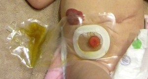
The bright pink bump on her abdomen is her stoma – a surgically created “opening” to her body. Technically her ostomy is called a jejunosotmy. The stoma has no nerve endings – so it doesn’t hurt her to touch it, nor does it hurt when she’s on her belly. The dark red circle on her abdomen above her stoma is one of her hemangiomas that we referred to in an earlier post.
Okay – here’s a quick anatomy lesson. The small intestine is made up of three structural parts: The duodenum (top), jejunum (middle), and the ileum (end). In Peanut’s first surgery (July 31st), they removed her ileum, which was about 10 inches (along with her appendix). It seems like a lot, but an adult female has somewhere around 23 feet of small intestine. Crazy! So Peanut’s jejunum is now the “end” of her small intestine. Her jejunum is surgically connected to the stoma on her abdomen so that her waste empties into the bag/pouch. In Peanut’s second surgery (Aug. 1st) she had most of her large intestine removed – the cecum and entire colon, leaving only a partial rectal stump and anal canal.
The scar:
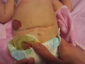
In the second surgery, the surgeons left her abdomen open and attached a “silo” under her skin exposing her remaining intestines. (If you Google “abdominal silo” you can get a better picture of what this is). With a silo, the doctors could literally peek at her intestines to keep their eye out for any signs of more NEC (Necrotizing Enterocolitis) developing. She remained like this for 7 days (on a paralytic so she didn’t move and pull out the silo). Thankfully, they did not have to remove any more intestine, so during her 3rd surgery (Aug. 8th) they removed the silo and closed her abdomen, leaving behind this lovely battle scar. This same incision site will be used for her next surgery. The surgeon assures us that he will make it look much “prettier” the next time around. I’m sure Peanut will appreciate that when she’s older, but it makes no difference to us since her daddy will never let her wear a 2-piece swimming suit anyway. 🙂
We hope this gives you a better understanding of some of the terms we use relating to Peanut’s condition in our posts. That concludes today’s “anatomy of a Peanut” lesson. Class dismissed!

Regardless of her anatomy she still is a sweetie pie. I love reading these updates about your family Jill.
Great job with the anatomy lesson, I’m super impressed. Despite her battle scars, she is a gorgeous, gorgeous child! Love her cute little face as she’s sprawled out on the towel!
I love to read your blog and get updates on peanut and your family, your anatomy lesson and pictures definately helps to better understand 🙂 She’s beautiful and looks like such a happy little girl!